


Page Under Maintenance
This page is currently under maintenance.
Please communicate at the hospital help desk for assistance.
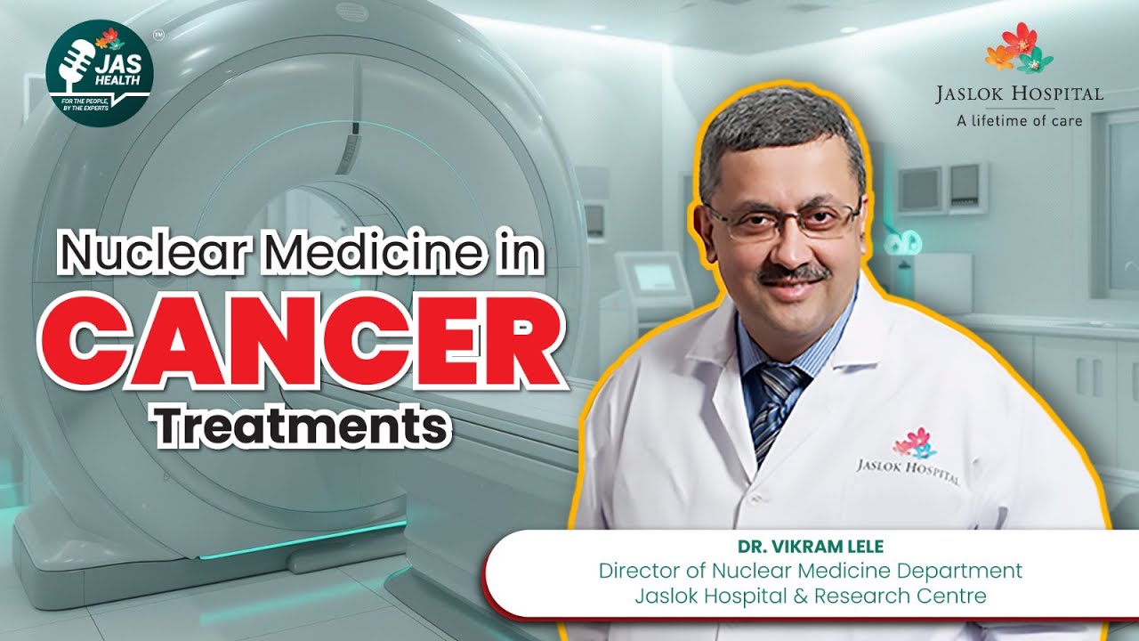
Cancer is a word that brings immediate fear to most people — but early detection dramatically improves treatment outcomes and prognosis. In this episode of Just Health for the People, a public health awareness initiative by Jaslok Hospital, Dr. Vikram Lele, Director of the Nuclear Medicine Department at Jaslok Hospital and Research Centre, Mumbai, explains how nuclear medicine plays a central role in detecting cancer early, tracking its spread, and increasingly, treating it directly.
Detecting cancer at an early stage makes treatment significantly easier and improves prognosis substantially. This is precisely why advanced detection technology — capable of identifying cancer before it spreads — plays such a critical role in modern oncology. Nuclear medicine is one of the key specialties enabling this early, precise detection.
Nuclear medicine uses radioisotopes, which are injected into the body intravenously. Specialized imaging equipment — including PET scanners and gamma cameras — then detects where these radioactive tracers accumulate in the body, revealing the location and spread of cancer.
Cancer cells are remarkably adaptive and mutate readily, but they share certain exploitable characteristics. One of the most useful is their metabolism: because cancer cells multiply extremely rapidly, they require significantly more energy than normal cells — and that energy comes primarily from glucose (sugar).
Dr. Lele explains that normal body tissues, like the brain and heart, also require glucose for energy. However, cancer cells demand roughly 15 times more glucose than normal body cells due to their rapid multiplication. By creating a radioactive version of glucose and injecting it into the body, doctors can track exactly where this glucose accumulates abnormally — revealing what's known as a "hot spot," a strong indicator of cancer activity.
This technique — injecting radioactive glucose and scanning the entire body from head to toe — is known as an FDG PET scan. Wherever abnormal glucose accumulation appears on the scan, it signals a likely cancerous area requiring further evaluation.
PET-CT combines two complementary imaging technologies:
Together, this combination makes PET-CT an exceptionally powerful tool for accurate cancer detection and staging.
Not all cancers are best detected through glucose metabolism alone. Many cancer cells express specific surface receptors that can be targeted with specialized tracers:
Prostate cancer cells express a protein called Prostate-Specific Membrane Antigen (PSMA) on their surface, which can be directly imaged using a specialized PSMA PET scan.
Neuroendocrine cancers — once considered rare and difficult to detect — express somatostatin receptors on their surface. Modern PET-CT technology can now identify these receptors with much greater ease, significantly improving detection of this previously elusive cancer type.
One of the most valuable applications of FDG PET-CT is early treatment response monitoring. Previously, chemotherapy would typically run for six months before a follow-up CT scan revealed whether it was effective — meaning patients could endure months of chemotherapy side effects only to learn the treatment wasn't working.
With FDG PET-CT, doctors can now assess treatment effectiveness after just one or two chemotherapy cycles. If a tumor that was previously showing aggressive glucose uptake suddenly shows little to no uptake after treatment, it's a strong sign the chemotherapy is working — allowing doctors to confirm or adjust treatment plans far earlier than before.
This approach is especially valuable in conditions like lymphoma (cancer of the lymph nodes), where PET scans are typically performed after every three chemotherapy cycles. If the scan shows good response, treatment continues; if not, doctors can switch to an alternative chemotherapy regimen promptly, rather than waiting for a full treatment course to conclude.
After successful treatment, regular follow-up PET scans help detect any recurrence at a very early stage — allowing for prompt intervention rather than discovering a relapse only once it has progressed significantly.
Not all cancers are equally aggressive. Some grow slowly and don't significantly utilize glucose, making them harder to detect via standard FDG PET scans. For these cases, doctors rely on the cancer's other unique characteristics — such as PSMA expression in prostate cancer — using targeted tracers like PSMA PET-CT to pinpoint where the disease has spread.
One of the most significant recent advances in nuclear medicine is theranostics — using the same targeting mechanism for both diagnosis and treatment. For example:
This theranostic approach is expanding rapidly, offering new hope for patients with prostate cancer, neuroendocrine tumors, and other cancer types that don't respond well to conventional chemotherapy, radiation, or surgery. Newer therapeutic isotopes — including Lutetium-PSMA, Lutetium-DOTA, and Lutetium-FAPI — allow doctors not only to diagnose these cancers with precision but to directly treat them, offering patients additional years of good-quality life even in advanced or treatment-resistant cases.
Nuclear medicine plays a valuable role across multiple stages of cancer care:
As these technologies continue to advance, nuclear medicine is increasingly central not just to diagnosing cancer, but to treating it with precision.
This article is based on an episode of Just Health for the People, a public health awareness initiative by Jaslok Hospital, featuring Dr. Vikram Lele, Director of Nuclear Medicine. Watch the full video here: Nuclear Medicine and Cancer Detection Explained by Dr. Vikram Lele
For consultation regarding cancer screening, PET-CT imaging, or radioisotope therapy, please reach out to Jaslok Hospital's Department of Nuclear Medicine.