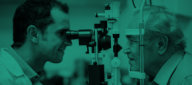
In a comprehensive eye exam, doctors use two primary tools to look at different "neighborhoods" of your eye. The Slit-Lamp is like a high-powered microscope for the front and middle sections, while Indirect Ophthalmoscopy is the specialized technique used to sweep the "back yard" (the peripheral retina) for hidden damage.
1. Slit-Lamp Examination (Biomicroscopy)
The slit-lamp is the centerpiece of the eye clinic. It combines a high-intensity light source that can be focused as a thin "slit" with a binocular microscope.
- What it looks at: It provides a 3D, magnified view of the eyelids, cornea, iris, lens, and the fluid-filled space in between. With special handheld lenses, it can also look at the central retina (macula) and optic nerve.
- The "Slit" Secret: By shining a thin sliver of light at an angle, the doctor can see the "optical sectioning" of the eye—allowing them to detect exactly which layer of the cornea or lens has a problem.
Common Symptoms for Testing
- Redness, itching, or discharge from the eyelids.
- The sensation of a "foreign body" (feeling like sand is in your eye).
- Blurred vision or seeing "halos" around lights (cataracts).
- Eye pain or sudden light sensitivity.
Associated Conditions
- Cataracts: Clouding of the eye's natural lens.
- Corneal Ulcers/Abrasions: Scratches or infections on the front window.
- Uveitis: Internal inflammation of the eye.
- Dry Eye Syndrome: Evaluating the "tear film" and tear ducts.
2. Indirect Ophthalmoscopy
While a slit-lamp is great for detail, its field of view is narrow. Indirect Ophthalmoscopy is used to get a wide-angle, panoramic view of the entire retina, including the far edges that are often missed by other tests.
- The Setup: The doctor wears a light on their head (the "headlight") and holds a large condensing lens (typically $+20D$ or $+28D$ power) in front of your eye.
- The View: The image the doctor sees is inverted and reversed, but it allows them to see the retina in 3D (stereoscopic) and through "hazy" conditions like a mild cataract or bleeding.
Common Symptoms for Testing
- Floaters: Seeing new "cobwebs" or spots drifting in your vision.
- Flashes: Seeing "lightning bolts" or sparks (a sign of retinal pulling).
- "Curtain" over vision: A sign of a potential retinal detachment.
- Diabetes Monitoring: Checking for leaky vessels in the peripheral retina.
Associated Conditions
- Retinal Detachment or Tears: Critical emergencies that require immediate surgery.
- Diabetic Retinopathy: Systemic damage to the blood vessels.
- Retinal Vein Occlusion: A "stroke" in the eye.
- Tumors: Checking for ocular melanoma or other growths.
3. Comparison at a Glance
|
Feature
|
Slit-Lamp Examination
|
Indirect Ophthalmoscopy
|
|
Primary Focus
|
Anterior Segment (Front)
|
Posterior Segment (Back/Retina)
|
|
Magnification
|
High ($10\times$ to $40\times$)
|
Low to Moderate ($2\times$ to $5\times$)
|
|
Field of View
|
Narrow and deep
|
Wide and panoramic
|
|
Patient Position
|
Sitting, chin in a rest
|
Sitting or Reclining
|
|
Stereo (3D) View
|
Yes
|
Yes
|
4. Pre and Post-Care Requirements
Pre-Care:
- Dilation: For a thorough exam (especially indirect), your eyes will be dilated with drops. These take 20–30 minutes to work and make your pupils large.
- Sensitivity: Dilation makes you very sensitive to light. Bring sunglasses to the appointment.
Post-Care:
- No Driving: Because dilation blurs your "near" vision and makes everything bright, you should not drive for 4–6 hours after the test.
- Reading Difficulty: You will find it hard to read fine print or use a smartphone for several hours until the drops wear off.
5. Hospitalization and Timeline
- Setting: These are outpatient procedures performed in the doctor's office.
- Procedure Time: * Slit-Lamp: 5 to 10 minutes.
- Indirect Ophthalmoscopy: 5 to 10 minutes (after dilation is complete).
- Total Stay: Usually 1 to 1.5 hours (mostly waiting for the dilation drops to work).
6. Benefits of These Procedures
- Early Detection: They can find "silent" issues like glaucoma or retinal holes before they cause permanent blindness.
- Non-Invasive: No part of the equipment (except the light and occasionally a numbing drop) needs to touch your eye.
- Real-Time Diagnosis: Most doctors can give you the results of these exams immediately after looking.